What is MINTS?
Multi-Scale Integrated Intelligent Interactive Sensing Consortium
The vision of MINTS is a holistic intelligent synergy of integrated targeted observations from multiple platforms on multiple spatial and temporal scales, together with state of the art compression and visualization systems for the distribution and comprehensive analysis of complete datasets.
MINTS is able to provide intelligent sensing solutions for your application, whether it concerns natural resources, environment and health, safety and security, or transportation and geospatial applications


Biometric Dashboard
The goal of this project was to process and visualize biometric signals in meaningful ways using modern visualization tooling.
- Visualize Electrical Activity from the Brain Visualizing Electroencephologram (EEG) signals helps researchers identify brain processes related to brain disorder, social interaction and anxiety, perception and intuition, etc.
- Visualize Heart Activity and calculate Heart Rate Variability Visualizing heart activity using Electrocardiogram (ECG) signals and calculating Heart Rate Variability (HRV) can help provide solid insights as to whether or not an individual is generally healthy.
- Calculate and Display Respiration Rate Using ECG signals, respiration rate can be accurately calculated. This measure can be used to detect abnormalities caused by illness as well as confirm overall health.
- Provide a measure for Electrodermal Activity Also known as Galvanic Skin Response (GSR). It measures skin conductance and can help identify the intensity of emotional arousal and cognitive load.
- Display Oxygen Saturation Levels Oxygen saturation levels are measured by finding peripheral capillary oxygen saturation (SpO2). SpO2 indicates how much oxygen is in the blood and can be used to determine how efficiently it is being carried to rest of the body.
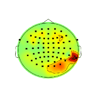
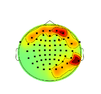
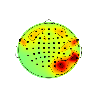
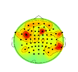
EEG Module
What does the EEG Module show?
Electroencephologram (EEG) evaluates brain activity. Electrodes are placed on the scalp and measure the electrical impulses that occur in the brain and record the results. The results are in the form of wave patterns that can be evaluated at different frequency bands. The goal for the EEG visualization is to plot the heatmaps for the delta band, theta band, alpha band, and total band, and show the electrical activity that is being picked up by the different sensors.
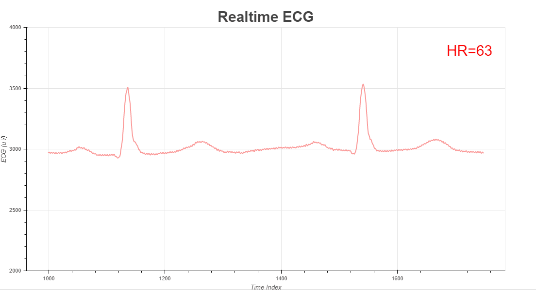
ECG Module
What does it show?
An ECG can be analyzed by studying the components of the waveform. The first initial upward tracing is the p wave, which indicates atrial contraction. The QRS complex, which begins with a small downward deflection,Q, followed by an upward deflection, which is called an R peak, and then a downward deflection S. The QRS complex indicates ventricular depolarization and contraction. Finally, the T wave, which is normally a smaller upwards waveform, representing ventricular repolarization.
Text Visualizations
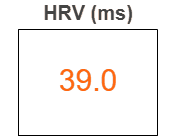
HRV
The HRV module leverages open source QRS detection code that uses the Pan-Tompkins QRS peak detection algorithm. The algorithm calculates beat-to-beat (RR) intervals and uses these intervals to calculate HRV values. Calculation is done for the rMSSD, SDNN, and SDSD, but the visualization shows the rMSSD value.
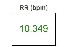
RR
With the RR interval calculated from the HRV module, the RR module calculates the respiration rate using one of two methods: Welch’s (default) and Fourier transformation. Welch’s alrogithm gives the number of seconds per breathing cycle (in Hz) and this number is then used to calculate breaths per minute.
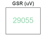
GSR
Skin conductance is captured using skin electrodes which are easy to apply. GSR devices typically consist of two electrodes, an amplifier (to boost signal amplitude), and a digitizer (to transfer the analog raw signal into binary data streams).
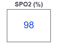
Sp02
Sp02 is measured using pulse oximeters that use light sensors to record how much blood is carrying oxygen and how much blood is not. Oxygen-saturated hemoglobin is darker than non-oxygen saturated hemoglobin, which allows the pulse oximeter to detect minute variations in the blood and translate that into a reading.